Surgical Success Rates Go Up In Smoke

blog post photo

If You Needed Another Reason to Stop Smoking, Here It Is…

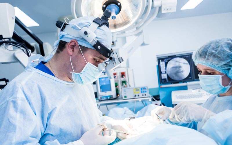
In medical research over the last few years, it has been proven conclusively that not only do smokers have a higher incidence of back pain; but when they do undergo surgery for back problems, they have a 15% higher failure rate. This appears to be related to the fact that the nicotine in the cigarette smoke constricts the small blood vessels which are critical to healing at the surgical site, especially when a fusion is being performed.

Smoking Constricts Blood Flow and Interferes With Healing

A fusion is when bone is moved from one portion of the body to another to create stability in that area of the body. Smoking greatly interferes with the ability of bone to heal. In fact, it has been shown that smoking may prevent some type of fractures from healing altogether. Instead of employing expensive and elaborate medical technology such as electrical stimulator and bone producing enzymes; a patient can get healing to occur simply by discontinuing smoking.

Smoking Cessation Solutions Are Available

At Sierra Regional Spine Institute we understand that it is not easy to discontinue the smoking habit. We make available to our patients a variety of specialized smoking cessation programs. These programs may include the use of medications, behavior modification, acupuncture and counseling. In a recent international spine meeting, an esteemed professor of orthopedic spinal surgery, Dr. Glen Rechtine, said that the most important thing that a doctor can do for any patient coming through his office door is to get them to stop smoking. If the threat of dying a miserable death from lung cancer was not enough to motivate you to stop smoking, then perhaps the prospect of leading a life in excruciating back pain will be the necessary motivation. We are here to help; talk to us about this serious problem.

Article written for “We’ve Got Your Back Magazine”

By Dr. James Rappaport
Accessibility Toolbar

decoration

Artificial Disc Replacement

At Sierra Regional Spine Institute, we believe that the future is restoration of anatomy and function through motion. The artificial disc replacement is the answer to that problem. Sierra Regional Spine Institute continues to be a leader in Nevada when disc replacement surgery is needed.
It is a surgical procedure in which degenerated intervertebral discs in the spinal column are replaced with artificial disc implants in the lumbar or cervical spine.

The procedure is used to treat chronic, severe low back pain and cervical pain resulting from degenerative disc disease.

Artificial disc replacement has been developed as an alternative to spinal fusion, with the goal of pain reduction or elimination, while still allowing motion throughout the spine. Another possible benefit is the prevention of premature breakdown in adjacent levels of the spine, a potential risk in fusion surgeries.

Lorem ipsum dolor sit amet consectetur adipscing elit.

decoration

Revision Spine Surgery

Revision spine surgery is a surgery procedure performed in certain patients to correct the problems of earlier spine surgery. Revision surgery is only when working with patients who experience chronic pain or any worsening symptoms even after the initial surgery.
Other factors that can indicate the need for revision spine surgery include:

Lorem ipsum dolor sit amet consectetur adipscing elit.

decoration

Decompression

At Sierra Regional Spine Institute we use Decompression in a few ways to treat and make the process of recovery smoother for people with fractures: Microdiscectomy, Microendoscopic Laminectomy, Minimally Invasive Cervical Foraminotomy (MICF), Vertebroplasty and Kyphoplasty.

Microdiscectomy

Microdiscectomy, also called microlumbar discectomy (MLD), is a very common MIS decompression procedure performed in patients with a symptomatic lumbar herniated disc. The operation consists of removing the portion of the intervertebral disc that is herniated and compressing a spinal nerve root.

Minimally Invasive Cervical Foraminotomy

This is a MIS cervical foraminotomy decompression procedure that enlarges the space in which a spinal nerve root exits the cervical spinal canal (intervertebral foramen). This narrowing can be caused by a herniated disc, bone spurs, thickened ligaments or joints, which may result in pinched nerves.

Vertebroplasty

Vertebroplasty for the treatment of vertebral compression fractures (VCFs) was introduced in the United States in the early 1990s. The procedure is usually done on an outpatient basis, although some patients stay in the hospital overnight. The procedure may be performed with a local anesthetic and intravenous sedation or general anesthesia.

Using x-ray guidance, a small needle containing specially formulated acrylic bone cement is injected into the collapsed vertebra. The cement hardens within minutes, strengthening and stabilizing the fractured vertebra. Most experts believe that pain relief is achieved through mechanical support and stability provided by the bone cement.
Kyphoplasty
Kyphoplasty involves an added procedure performed before the cement is injected into the vertebra. First, two small incisions are made and a probe is placed into the vertebral space where the fracture is located.

The bone is drilled and one balloon (called a bone tamp) is inserted on each side. The two balloons are then inflated with contrast medium (which are visualized using image guidance x-rays) until they expand to the desired height and removed. The spaces created by the balloons are then filled with the cement. Kyphoplasty has the added benefit of restoring height to the spine.

Lorem ipsum dolor sit amet consectetur adipscing elit.

decoration

Cervical Fusion

Cervical Fusion is an operation that creates a solid union between two or more vertebrae in the upper spine (neck) area. This procedure may assist in strengthening and stabilizing the spine and may thereby help to alleviate severe and chronic back pain.
The best clinical results are generally achieved in single-level fusion, although fusion at two levels may be performed in properly selected patients.

Bone grafts may be taken from the hip or from another bone in the same patient (autograft) or from a bone bank (allograft). Bone graft extenders and bone morphogenetic proteins (hormones that cause bone to grow inside the body) can also be used to reduce or eliminate the need for bone grafts.

Fusion sometimes involves the use of supplemental hardware (instrumentation) such as plates, screws, and cages. This fusing of the bone graft with the bones of the spine will provide a permanent union between those bones. Once that occurs, the hardware is no longer needed, but most patients prefer to leave the hardware in place rather than go through another surgery to remove it.

Fusion can sometimes be performed via smaller incisions through MIS techniques. The use of advanced fluoroscopy and endoscopy has improved the accuracy of incisions and hardware placement, minimizing tissue trauma while enabling an MIS approach.

Lorem ipsum dolor sit amet consectetur adipscing elit.

decoration

Lumbar Fusion

This is a MIS technique that is performed in patients with refractory mechanical low back and radicular pain associated with spondylolisthesis, degenerative disc disease, and recurrent disc herniation. The procedure is performed from the back (posterior) with the patient on his or her stomach.

Using x-ray guidance, two 2.5-cm incisions are made on either side of the lower back. The muscles are gradually dilated and tubular retractors inserted to allow access to the affected area of the lumbar spine.

The lamina is removed to allow visualization of the nerve roots. The disc material is removed from the spine and replaced with a bone graft and structural support from a cage made of bone, titanium, carbon-fiber, or a polymer, followed by rod and screw placement.

The tubular retractors are removed, allowing the dilated muscles to come back together, and the incisions are closed. This procedure typically takes about 3 to 3 1/2 hours to perform.

Outcome & Benefits

Pioneers of both surgical and non-surgical techniques

decoration

Minimally Invasive Spine Surgery

MIS was first performed in the 80s but has recently seen rapid advances. Technological advances have enabled surgeons to expand patient selection and treat an array of spinal disorders, such as degenerative disc disease, herniated disc, fractures, tumors, infections, instability, and deformity.
This can result in quicker recovery, decreased operative blood loss, and speedier patient return to normal function. In some MIS approaches, also called, “keyhole surgeries,” surgeons use a tiny endoscope with a camera on the end, which is inserted through a small incision in the skin. The camera provides surgeons with an inside view, enabling surgical access to the affected area of the spine.

Benefits of Minimally Invasive Surgery

Lorem ipsum dolor sit amet consectetur adipscing elit.